A group of researchers from the I3A, at the University of Zaragoza, work from Engineering and Mathematics to know the functioning of the heart, see how its activity is when it is healthy and how it changes when a heart attack occurs, provide clinical specialists with the tools that they need to improve patient care.
They do it through computation in cardiology, with the creation of a virtual heart that allows the computer to reproduce the electrical activity of a real heart. Their line of research advances towards a simpler methodology. Until now, computer simulation required the construction of a geometry and the creation of a mesh, joining different points of that virtual heart. This system was not so straightforwardly applicable to the clinical routine, as extensive engineering knowledge was needed. Now they have created a new methodology that facilitates that application because it easily translates an image into a computational model and, therefore, it can be easier to interpret in the hospital environment.
It is an innovative advance in this field and its work has already been recognized in the Computing in Cardiology (CinC) Conference held recently and where they have received the “Maastricht Simulation Award (MSA)”.
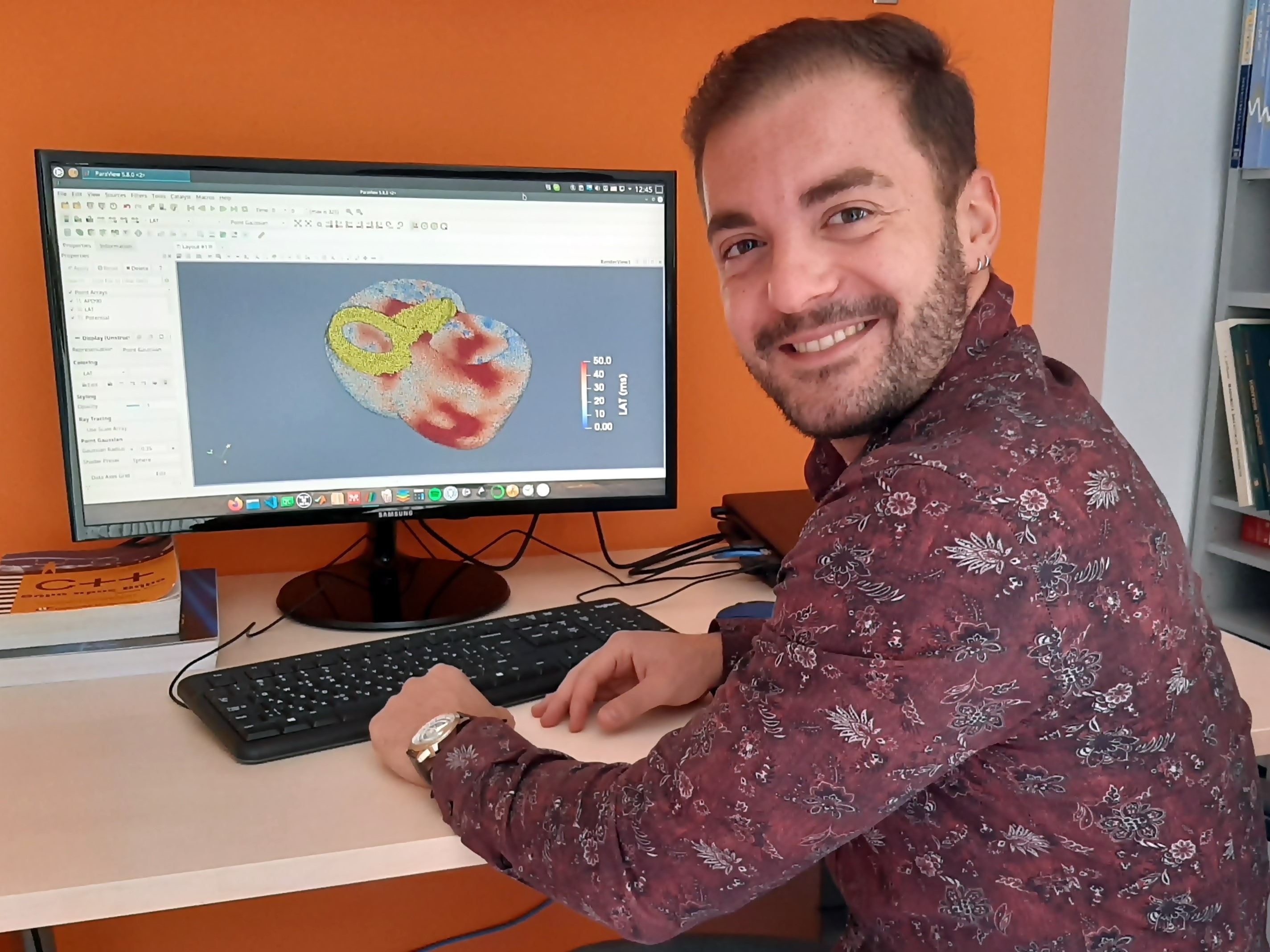
Konstantinos Mountris is part of the BSiCoS research group, at the I3A, he was in charge of presenting this work where he makes a simulation of myocardial infarction. He reproduces the functioning of the heart in its real situation on a computer, but for that, knowledge of the anatomy and electrophysiology of that vital organ is needed.
Until now, this group of researchers started from a clinical image and reconstructed a geometry that they had to divide into small pieces and establish their connection. With this new methodology, this is not necessary, you do not have to build the virtual heart by connecting those small parts to see how it works, but they start from a set of points generated according to the image itself. A model is built automatically and they are able to see heart activity.
This methodology that unites Engineering and Mathematics “is applicable to different pathologies of the heart, but in the work that we present it had been tested against myocardial infarction. Our idea is to test the electrical activity of the heart that has suffered a heart attack”, explains Konstantinos Mountris, but they also test the activity in a healthy heart.
 Transferring the image of a damaged heart to the computer simulation allows us to check how its activity will be from now on, how it will behave and this could help clinicians in their diagnosis, application of treatments and decision-making.
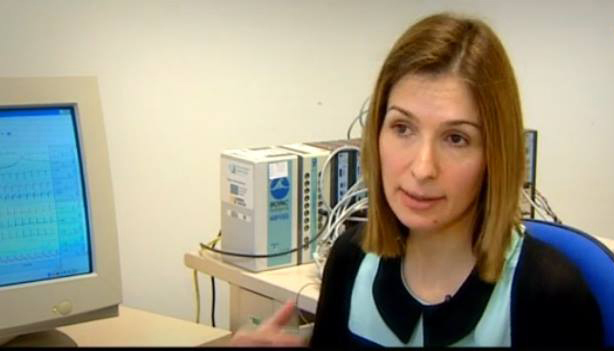
It is a method with a high mathematical and engineering load but with a wide clinical application, "these are algorithms that could tranferred to the clinic and obtain a result from the image that doctors have", highlights Esther Pueyo, principal investigator of the European Modelage Project, in which the work that has just been internationally recognized is framed.
This line of research proposes a method that has different applications, from surgeries to diagnostic tests or treatments. A mathematical model that reproduces the functioning of a healthy heart or a heart with areas affected by an arrhythmia or a heart attack and can be adapted to each patient.
Modelage is a European Project that tries to understand the aging rhythms of the heart and develop patterns that help prevent arrhythmias. It is led by Esther Pueyo, researcher at the BSICoS group at I3A (University of Zaragoza). It was selected within the first Starting Grant call of the Horizon 2020 program of the European Union in which more than 3,200 proposals competed. (More info)
Work Presentation in the Computing in Cardiology Conference . See video
